If you had to decide to exclusively use a rotary telephone or a smart phone, which would you reach for? The answer for most people is quite simple, and this is quickly becoming the case for orthopedic surgeons as well. The availability of 3D-CT software is making it easier for surgeons who want to utilize such advancements for preoperative planning, and studies continue to show that this is good for just about everyone. One particular study dives into the benefits of preoperative planning via various 3D-CT applications.
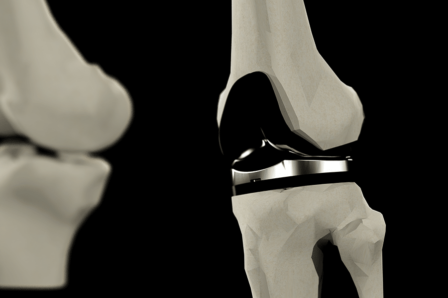
The standard method for planning knee and hip arthroplasties is by superimposing template films overtop of plain radiographs to determine the likely size and position of the implant. This process, while still widely used, has the potential to lack accuracy when making decisions about three-dimensional bony structures based on two-dimensional data. Within a 3D-CT planning software, the patient’s CT images are converted into a three-dimensional representation. The surgeon can then more easily make adjustments to implant sizing and orientation with a better general idea of how to achieve the best fit. This has been shown to reduce the intraoperative guesswork when selecting implant size for both acetabular and femoral components. In general, the authors of this study suggest that surgical planning via 3D-CT enhances surgical precision of joint replacement surgery as well as helps surgeons anticipate potential complications such as femoral fractures or leg length inequality.
The potential benefits of 3D-CT joint replacement planning from a patient perspective is pretty easy to see. Your surgeon has access to a higher degree of visual fidelity and thus is able to plan a more accurate procedure. With any new medical technique, there needs to be more randomized controlled trials examining the effectiveness of this at long-term intervals, before any conclusive statements are made. However, the evidence reported at a five year follow-up showed good clinical outcomes with successful leg length restoration and hip center of rotation, all with a low rate of complications.
In summary, 3D-CT preoperative planning for total joint arthroplasties is a huge step in the right direction toward more accurate, more efficient, and more functional procedures. There are always going to be a few barriers to converting a medical practice in order to accommodate 3D-CT capabilities as standard operating procedures. However, the evidence suggests that it’s both worthwhile for surgeons and their patients.
At Kinomatic, we fully recognize the significance that 3D-CT models can offer the orthopedic industry. That is why we go one step further by introducing virtual reality. As we’ve discussed, 3D models offer much more flexibility with preoperative planning than 2D radiographs, however, those 3D images are still viewed on 2D monitors. Virtual reality allows a surgeon to see true patient imaging in full, stereoscopic 3D. So, if we go back to our phone analogy, and equate 2D templating from radiographs to old, rotary telephones, we would then say that 3D templating on a 2D screen would be the world’s first cordless phones. And if 3D templating on a 2D screen is the first cordless phone, then Kinomatic Custom Surgical Planning in virtual reality would be the iPhone 13 with 5G.
If you’d like to know more about how Kinomatic leverages 3D models and virtual reality, there is some great information on our website.
If you’d like to read more about the benefits of 3D-CT for total joint arthroplasty read the full article below:
http://www.opnews.com/2017/10/3d-ct-better-map-hip-surgery/14077
Authors:
Alister Hart – Consultant Orthopaedic Surgeon, Royal National Orthopaedic Hospital (RNOH), & Chair of Academic Clinical Orthopaedics, University College London (UCL), Jia Zhe Su – Medical Student, University College London (UCL), Mr Johann Henckel – Hon Fellow, Royal National Orthopaedic Hospital (RNOH), Anna Di Laura – Biomedical Engineer & PhD Student, University College London (UCL), Klaus Schlüter-Brust – Chefarzt und Leitung des Endoprothetik Zentrum der Maximalversorgung EPZ (max) am SFH bei St. Franziskus Hospital Köln, Germany
