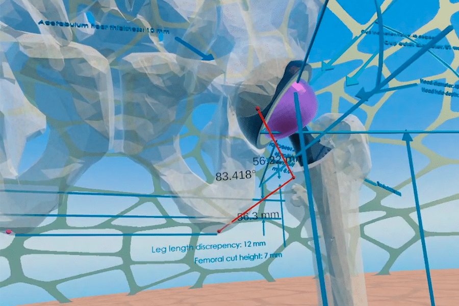
Five experienced otologic surgeons have made an even stronger case for preoperative planning in virtual reality compared to conventional cross-sectional imaging. Consumer grade virtual reality headsets are more available and approachable than ever before. The immersiveness that VR provides lends itself well to a plethora of industries, healthcare being a continuously growing one. Specifically, as this study points out, the use of VR during preoperative planning has the capacity to become the dominant method of surgical planning due to fewer limitations and a fully 3-dimensional view of patient anatomy.
Patient imaging is a key aspect of planning a successful surgery. For complex procedures and for the surgeons responsible for this study preoperative imaging is mostly performed with computed tomography (CT) and magnetic resonance imaging (MRI). Despite the advances in preoperative imaging, these images are still viewed in 2D cross sections thus forcing the surgeon to map out an understanding of complex three-dimensional anatomy. Virtual reality breaks through this barrier by allowing the user to view a computer generated environment in full, stereoscopic 3D. When you combine the immersive environment with motion tracking controllers, you achieve a level of flexibility while viewing multidimensional anatomy that is currently not possible with conventional cross-sectional 2D images.
The method for this study involved five normal cadaver temporal bones and added multiple fiducial markers at predetermined locations. These markers were then measured physically with a standard Vernier caliper under a microscope. The distances between the markers were then compared with the distances measured in the VR application and the conventional 2D interface.
The five surgeons who volunteered for this study had an average operating experience of 16.5 years. None of the surgeons had any prior experience with VR. When surveyed about the experience of measuring the temporal bone structures the average, overall responses significantly favored the VR method. Measures such as the appearance of anatomical structures, hand-eye coordination ability, and overall understanding of the surgical site were considerably preferred in VR.
Given the experience of the five surgeons in the study, there were not significant differences in the measurements of the fiducials between the VR method and cross section method with an average of only 1.9%. However, there were more cases of misidentification of fiducials in the cross-sectional viewing.
What this study shows is that using virtual reality as a preoperative planning tool can provide surgeons with an accurate, reliable, and in these cases, preferable method for surgical planning when compared against conventional cross-sectional viewing.
As computing software and virtual reality hardware advance one could make a compelling case that VR surgical planning will become the new standard for most surgical cases. VR offers a much more understandable and tangible way to view and assess the complexities of the human body.
To read more about how Kinomatic is utilizing VR for preoperative surgical planning, follow the link below:
https://www.kinomatic.com/how-it-works/
Timonen, T., Iso-Mustajärvi, M., Linder, P. et al. Virtual reality improves the accuracy of simulated preoperative planning in temporal bones: a feasibility and validation study. Eur Arch Otorhinolaryngol 278, 2795–2806 (2021). https://doi.org/10.1007/s00405-020-06360-6
